
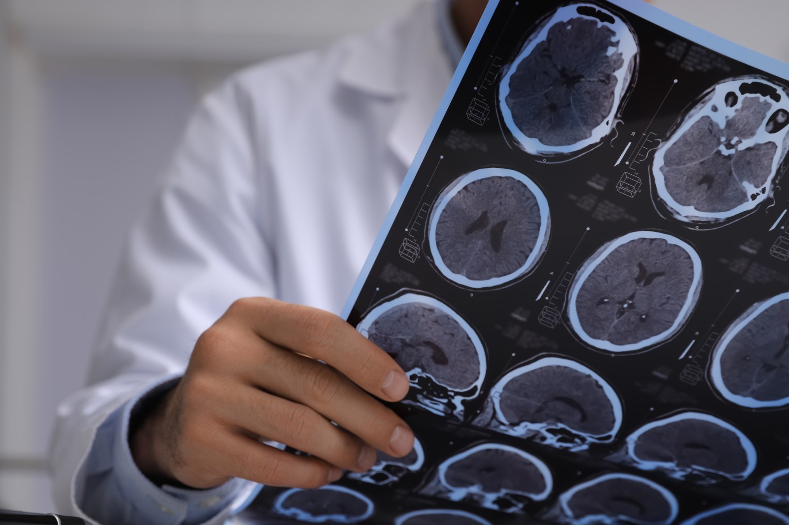
New research indicates that changes in the brain linked to Alzheimer’s disease can occur more than seven years before conventional imaging methods can detect the condition. Traditionally, the appearance of amyloid plaques on amyloid-PET scans has been considered the initial detectable sign of Alzheimer’s. Current findings suggest that structural changes visible on MRI scans may emerge earlier, implying that the disease process initiates much sooner than previously recognized.
A study tracked cognitively healthy older adults without signs of memory or cognitive issues. Participants underwent repeated MRI brain scans over nearly 20 years. Some participants eventually developed amyloid plaques, while others did not. Researchers scrutinized the point at which plaques became detectable on PET scans and analyzed MRI scans from the preceding decade.
The study found that individuals who later developed plaques already exhibited measurable brain structure changes years in advance. This observation suggests that such changes appear at least seven years prior to the visibility of amyloid plaques on scans. These findings represent the earliest brain-imaging signal related to Alzheimer’s detected so far.
The study, published in Nature Neuroscience, prompts questions about how scientists define the onset of Alzheimer’s. If changes linked to Alzheimer’s occur before plaques emerge on scans, existing methods may overlook a significant segment of the disease process.
Researchers proposed two potential explanations. First, biological processes associated with Alzheimer’s might already be affecting the brain before plaque development. These processes may contribute to or occur alongside early plaque-related changes undetectable by current imaging techniques. Alternatively, different biological mechanisms could initiate brain changes before amyloid accumulation begins. This understanding could broaden the search for new treatment targets beyond amyloid plaques.
Dr. Lucy Hooper, a private GP and co-founder of Coyne Medical, believes the findings could change perceptions about intervention timing. “Knowing that Alzheimer’s evolves over years indicates a longer window for prevention or delay,” she told Newsweek. Hooper referenced prior research showcasing that cerebrospinal fluid and blood changes are detectable decades before brain scans reveal amyloid buildup, reinforcing the idea that Alzheimer’s commences long before traditional imaging identifies it.
Hooper explained that several biological processes might clarify why structural brain changes appear before plaques. She stated, “Amyloid in the brain is detected by PET scans, a specialized brain scan. Yet numerous changes can occur before amyloid is visible on a scan.” Soluble forms of amyloid may impair delicate brain connections before becoming visible as plaques. Factors like inflammation, immune cell changes, blood vessel alterations, and disruptions to the brain’s glymphatic system might occur separately from plaque accumulation.
The findings could also impact future diagnosis and treatment improvements. “Understanding biology thoroughly enhances Alzheimer’s diagnosis and prevention,” Hooper said. She mentioned that blood testing for plasma p-tau217 is already used clinically to expedite Alzheimer’s diagnoses, despite not being a faultless screening tool due to false positive and negative results. Better biological markers could aid in developing new therapies aimed at reducing or preventing Alzheimer’s disease.
While most current treatments target amyloid, researchers increasingly explore alternatives focusing on inflammation, metabolic changes, and the glymphatic system.
Reference: James M. Roe, et al. Cortical thickness changes precede high levels of amyloid by at least 7 years. Nature Neuroscience, 2026; DOI: 10.1038/s41593-026-02363-4
For inquiries, contact Newsweek editors Kara Dolman and Gray R. Thomas.